Showing posts with label Respiratory System. Show all posts

Wednesday, January 26, 2022

A 75-year-old woman with a 50-pack-year history of cigarette smoking presents to her primary care physician (PCP) with 10 years of progressive dyspnea on exertion


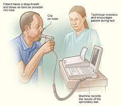
A 75-year-old woman with a 50-pack-year history of cigarette smoking presents to her primary care physician (PCP) with 10 years of progressive dyspnea on exertion. She notes episodic wheezing and experiences a “chest cold” about two times per year. Her examination reveals distant breath sounds to auscultation and hyper-resonant chest to percussion, with otherwise unremarkable examination. A CXR demonstrates hyperinflation with flattening of the bilateral hemidiaphragms. Pulmonary function tests (PFTs) reveal a forced expiratory volume (FEV1) of 50% predicted, forced vital capacity (FVC) of 70% predicted, and FEV1/FVC of 0.50. There is no response to bronchodilator.

What pattern best describes her PFT results?

A. Cannot determine without more information

B. Normal

C. Obstructive

D. Restrictive

Answer:

Tuesday, January 25, 2022

A 75-year-old man with a history of hypertension and peptic ulcer disease presents with dyspnea on exertion

 A 75-year-old man with a history of hypertension and peptic ulcer disease presents with dyspnea on exertion to the emergency department (ED). He notes black-colored stools for the past 2 weeks. His initial examination is notable for conjunctival pallor, clear lung fields without rales, wheezing, or rhonchi, as well as tachycardia with a regular rhythm, normal s1 and s2, and without murmurs, rubs, or gallops. Initial laboratory workup reveals a hemoglobin of 5.5 g/dL, reduced from a baseline of 12 g/dL on routine outpatient laboratory testing from 3 months prior. His basic metabolic panel, arterial blood gas (ABG), and lactateare all within normal limits. A chest radiograph (CXR) is normal.

What is the mechanism of this man’s dyspnea on exertion?

A. Decreased cardiac output

B. Decreased oxygen delivery

C. Decreased systemic vascular resistance

D. High-output heart failure

E. Pulmonary edema

Answer:

Wednesday, July 12, 2017

Anatomical Dead Space


The anatomical dead space may be used to calculate alveolar ventilation by subtracting it from the tidal volume and multiplying the result by the respiratory rate.

What would you expect the normal anatomical dead space to be in a healthy adult male?
A) 50 ml
B) 150 ml
C) 250 ml
D) 350 ml
E) 450 ml

Answer:

Saturday, July 1, 2017

Regarding Atypical Mycobacteria Affecting a Patient With Bronchiectasis...


A 68-yr-old man with bronchiectasis is found to have Acid fast bacilli in his sputum. The microbiology report suggests this may be an opportunistic or atypical mycobacterium.

Which of the following is the least likely infectious agent?
A) Mycobacterium kansasii
B) Mycobacterium malmoense
C) Mycobacterium xenopi
D) Mycobacterium leprae
E) Mycobacterium avium intracellulare


Answer:

Friday, June 23, 2017

A 26 Year Old Woman presents To Her General Physician With Persistent Cough


A 26-year-old teacher has consulted her general practitioner (GP) for her persistent cough. She wants to have a second course of antibiotics because an initial course of amoxicillin made no difference. The cough has troubled her for 3 months, since she moved to a new school. The cough is now disturbing her sleep and making her tired during the day. She teaches games, and the cough is troublesome when going out to the playground and when jogging.
In her medical history she had her appendix removed 3 years ago. She had her tonsils removed as a child and was said to have recurrent episodes of bronchitis between the ages of 3 and 6 years.
She has never smoked and takes no medication other than an oral contraceptive. Her parents are alive, and well and she has two brothers, one of whom has hay fever.

On Examination
The respiratory rate is 18/min. Her chest is clear, and there are no abnormalities in the nose or pharynx or the cardiovascular, respiratory or nervous systems.

Investigations:
• Chest X-ray is reported as normal.
• Spirometry is carried out at the clinic, and she is asked to record her peak flow rate at home, the best of three readings every morning and every evening for 2 weeks. Spirometry results are as follows:
FEV1 (L) = 3.9   ( predicted = 3.6–4.2)
FVC (L) = 5.0     ( predicted =  4.5–5.4)
FER (FEV1/FVC) (%) = 78   ( predicted = 75–80)
PEF (L/min)  =  470   ( predicted = 440–540)

Peak flow recording at home showed diurnal variations.

Questions
• What is your interpretation of these findings?
• What do you think is the likely diagnosis, and what would be appropriate treatment?

Answers and Case Discussion:

Thursday, June 22, 2017

Diagnosis for a 60 year-old man who is referred following a Chest X ray that suggests interstitial lung disease.


A 60 year-old man is referred following a Chest X ray that suggests interstitial lung disease. You proceed to bronchoscopy with transbronchial lung biopsy to try and make a definitive histological diagnosis.
Which of the following is the least likely diagnosis to be confirmed in this way?
A. Cryptogenic fibrosing alveolitis (CFA)
B. Sarcoidosis
C. Extrinsic allergic alveolitis
D. Cryptogenic organising pneumonia (COP)
E. Lymphangitis carcinomatosa

Answer;

Wednesday, June 14, 2017

A 20-year-old female college student presents with a 5-day history of cough and low-grade fever ...


A 20-year-old female college student presents with a 5-day history of cough, low-grade fever (temperature 100°F), sore throat, and coryza.
On examination, there is mild conjunctivitis and pharyngitis. Tympanic membranes are inflamed, and one bullous lesion is seen. Chest exam shows few basilar rales.
Laboratory findings are as follows:
Hct: 38
WBC: 12,000/μL
Lymphocytes: 50%
Mean corpuscular volume (MCV): 83 nL
Reticulocytes: 3% of red cells
CXR: bilateral patchy lower lobe infiltrates


The sputum Gram stain is likely to show
a. Gram-positive diplococci
b. Tiny gram-negative coccobacilli
c. White blood cells without organisms
d. Acid-fast bacilli

Answer:

A 19-year-old boy has a history of repeated chest infections.- Case Study

History
A 19-year-old boy has a history of repeated chest infections. He had problems with a cough and sputum production in the first 2 years of life and was labelled as bronchitic. Over the next 14 years he was often ‘chesty’ and had spent 4–5 weeks a year away from school. Over the past 2 years he has developed more problems and was admitted to hospital on three occasions with cough and purulent sputum. On the first two occasions, Haemophilus influenzae was grown on culture of the sputum, and on the last occasion 2 months previously, Pseudomonas aeruginosa was isolated from the sputum at the time of admission to hospital. He is still coughing up sputum. Although he has largely recovered from the infection, his mother is worried and asked for a further sputum sample to be sent off. The report has come back from the microbiology laboratory showing that there is a scanty growth of Pseudomonas on culture of the sputum.
There is no family history of any chest disease. Routine questioning shows that his appetite is reasonable, micturition is normal and his bowels tend to be irregular.

Examination
On examination he is thin, weighing 48 kg, and is 1.6 m (5 ft 6 in) tall.
• The only finding in the chest is of a few inspiratory crackles over the upper zones of both lungs. Cardiovascular and abdominal examination is normal.

The chest X-ray is shown below:

Questions
• What does the X-ray show?
• What is the most likely diagnosis?
• What investigations should be performed?

Answers And Discussion:

Tuesday, June 13, 2017

A 36-yr-old woman with systemic sclerosis develops breathlessness on exertion. ...


A 36-yr-old woman with systemic sclerosis develops breathlessness on exertion. Her pulmonary function tests show normal spirometry but a decreased gas transfer factor (TLCO, transfer factor for carbon monoxide) and transfer coefficient (KCO).

Which of the following is the most likely explanation for this abnormality?
A. Fibrosing alveolitis
B. Pulmonary vascular disease
C. Severe thoracic skin thickening
D. Pleural involvement
E. Respiratory muscle weakness

Answer:

Friday, June 9, 2017

A 44 year old man who keeps pigeons presents with pneumonia....


A 44-yr-old man is admitted with right-sided pneumonia. According to him he has been unwell for three to four days with malaise, fever, cough and muscular pain. He also has a rash on his abdomen and neck pain.
He was previously fit and has not traveled abroad. He is a plumber and also keeps pigeons. According to his wife, two of his favorite pigeons died two weeks ago.

Which organism would you be suspicious of as being responsible for his pneumonia?
A. Streptococcus pneumonia
B. Legionella pneumophila
C. Coxiella burnetii
D. Chlamydia psittaci
E. Mycoplasma pneumoniae

Answer:

Wednesday, June 7, 2017

Which of the following is a recognized treatment for complications of cystic fibrosis?


Which of the following is a recognized treatment for complications of cystic fibrosis?

A) DNAase to assist in reinflating collapsed lung segments.
B) Rectal pull-through and anastamosis for rectal prolapse.
C) Pancreatic transplant for diabetes mellitus.
D) Nebulised tobramycin for pseudomonas colonisation of the lower respiratory tract.
E) Hypotonic saline drinks for hypernatraemic dehydration.

Answer:

Saturday, June 3, 2017

A 45 Year old woman a known case of SLE Presents With Worsening Dyspnoea...


A 45-yr -old woman known to suffer from (SLE) is referred to the chest clinic with a history of worsening dyspnoea. She is on a maintenance dose of 15 mg of prednisolone. She has never smoked, and according to her she developed an unproductive cough and dyspnoea three months ago. There is no history of fever, night sweats or weight loss. Her doctor has tried various types of inhalers with no benefit.  A PEFR diary shows no nocturnal dips.
A Chest X Ray shows a reticulonodular pattern.
Examination is unremarkable apart from the skin changes of SLE.

What is the most likely Diagnosis?
A. Asthma
B. Bronchiectasis
C. Bronchiolitis obliterans
D. TB
E. Fungal infection

Answer:

Saturday, May 27, 2017

Superior vena cava obstruction - Case Study


A 65 years old man  who is a smoker with no h/o hypertension or diabetes presented with frequent episodes of difficult breathing and cough for last 6 months. Cough is present throughout the day and night and is is usually dry, sometimes associated with scanty mucoid expectoration. He also noticed small amount of blood with sputum several times. The patient also complains of loss of appetite and substantial weight loss for the same duration. There is no history of fever, chest pain or contact with TB patients.
For the last few days, he has noticed gradual swelling of the face and arms along with shortness of breath, which is more marked on exertion. The patient also complains of headache which is
aggravated by cough and movement of the head for the same duration. He denies any change of
voice, back pain, yellow coloration of urine and sclera. His bowel and bladder habits are normal.
He smokes about 30 sticks/day for 25 years.

On Examination: the chest movement during respiration were decreased on the right side, and the percussion note was dull in the right upper chest. On auscultation of the lung fields, there were decreased breath sounds on the right upper chest, and few crepitations were heard throughout.
Heart sounds were normal.
Fundoscopy shows dilated vessels, hemorrhage and exudates.
Other systemic examination was normal.

Provisional Diagnosis: Superior vena cava (SVC) obstruction

Q. What do you think the cause of SVC obstruction in this case?
According to the age of the patient in this case 65 years old so causes are:

  • bronchial carcinoma, 
  • lymphoma

In young or early age—common cause is lymphoma.

Q. Tell one single investigation which will help the diagnosis of SVC obstruction.
Chest X-ray (which may show bronchial carcinoma and lymphoma).

Q. What investigations should be done in SVC obstruction?
 As follows:
1. Chest X-ray
2. CBC, ESR
3. Sputum for malignant cells
4. CT or MRI of chest
5. Others (according to suspicion of cause or physical findings):

  •  If palpable lymph nodes FNAC or biopsy
  •  Bronchoscopy and mediastinoscopy, venography and occasionally thoracotomy may be needed
  •  Echocardiography in some cases.

Thursday, May 11, 2017

Pneumothorax - Long case study With Questions & Answers

Presenting Complains: 

  • Breathlessness for … days
  • Right sided chest pain for … days
  • Cough for … days.

History of present illness: According to the statement of the patient, he was alright … days back. Since then, he suddenly felt severe breathlessness associated with pain in right side of his chest.
Breathlessness is present in rest, more marked on lying on right side and also on lying flat. It is nonprogressive, not associated with wheeze and does not relieve by taking rest or drugs. He also complains of right sided chest pain, which is sharp and stabbing in nature, aggravated by deep breathing, coughing, with movement and lying on right side but no radiation. The patient also complains of cough with slight mucoid expectoration but no hemoptysis. There is no history of trauma or fever.
History of past illness: There is no history suggestive of COPD, or chronic bronchitis or bronchial
asthma. There is no previous attack of such illness.
Family history: Nothing contributory.
Socioeconomic history: He is a laborer, living in a slum area with poor sanitary facilities.
Personal history: He smokes 30 to 40 sticks/day for 20 years, but nonalcoholic.

General Physical Examination: 
  • The patient appears anxious and slightly dyspneic
  • Decubitus: patient prefers to lie on left lateral position
  • He is mildly anemic
  • No jaundice, cyanosis, edema, clubbing, koilonychia or leukonychia
  • No lymphadenopathy or thyromegaly
  • Respiratory rate: 34/min
  • BP: 110/80 mm Hg
  • Pulse: 108/min.
Respiratory System:(supposing right side)
- Inspection:
  • Restricted movement on right side of the chest
  • Intercostal spaces appear full.
- Palpation:
  • Trachea—deviated to the left
  • Apex beat—in left 6th intercostal space in anterior axillary line (shifted to left), normal in character
  • Vocal fremitus—reduced in right side but normal on the left side
  • Chest expansion—reduced on the right side of the chest.
 - Percussion: 
  • Hyper-resonance in right side (tell where), but normal on the left side
  • Upper border of the liver dullness—in the right 6th intercostal space in the midclavicular line.
 - Auscutation:
  • Breath sound—diminished (or absent) on the right side of the chest (tell up to which space), but vesicular on the left side.
  • Vocal resonance—diminished (or absent) on the right side of the chest (tell up to which space), but normal on the left side.
Examination of other systems reveals nothing abnormal.

1. What is your diagnosis?

Tuesday, May 9, 2017

A 65 Year Old Man Known Case Of Rheumatoid Arthritis develops A Pleural Effusion...


A 65-yr -old man with severe RA is admitted with a right pleural effusion. He has been
complaining of dyspnoea on exertion for the last three months. He has never smoked and has not
worked for over 20 yrs when he was diagnosed to be suffering from rheumatoid arthritis. Which
of the following is true?
A. Pleural effusions with RA occur in over 50% of pts
B. A glucose level in pleural fluid of < 1.6 mmol/l is characteristic of a rheumatoid pleural effusion
C. Pleural effusions associated with RA have low levels of cholesterol
D. The most appropriate Rx is chemical pleurodesis
E. Bilateral pleural effusions do not occur in RA

Answer:

Friday, May 5, 2017

Bronchial Carcinoma - Long Case Discussion With Questions & Answers.


Mr …, 52 years old, a clerk, normotensive, nondiabetic, smoker, hailing from …, presented with
frequent cough for … months, which is present throughout the day and night. The cough is usually
dry, sometimes associated with slight mucoid sputum expectoration. Occasionally, the patient noticed
streaks of frank blood with sputum during coughing. There is one episode of massive hemoptysis …
days back. For the last … days, he is also complaining of right sided chest pain, which becomes worse with deep inspiration, coughing and also on lying down on right side. The patient also complains of loss of appetite, substantial loss of weight, weakness and malaise for ... days. His bowel and bladder habits are normal. There is no history of fever, hoarseness of voice, difficulty in deglutition, contact with TB patients. He smokes about 25 sticks a day for the last 30 years. He used to take some cough syrup and occasionally antibiotics, the name of which he cannot mention.

Examination:
Respiratory System
(Supposing right sided)
Inspection:

  • Movement is restricted on right upper chest
  • There is radiation mark on the chest (if any).

Palpation:

  • Trachea is central in position
  • Apex beat is in left 5th intercostal space, just medial to the midclavicular line
  • Vocal fremitus is reduced (or absent) in right upper chest
  • Pleural rub is present (mention where).

Percussion:

  • Percussion note is dull in right upper chest (mention up to which space)
  • Upper border of the liver dullness is in the right 5th intercostal space in the midclavicular line
  • Cardiac dullness is normal.

Auscultation:

  • Breath sound is reduced (or absent)
  • Vocal resonance is reduced (or absent)
  • Few crepitations (may be present)
  • Pleural rub is present (mention where).

Examination of the other systems reveals no abnormalities.

1. What is your diagnosis? 
My diagnosis is bronchial carcinoma with metastasis (in right supracalvicular Lymph node).

2. Why bronchial carcinoma?
The patient is elderly and heavy smoker. There is history of cough with hemoptysis and marked loss
of weight. On examination, the patient is emaciated, has generalized clubbing with nicotine stain and
examination of chest shows evidence of mass lesion. All the features are suggestive of bronchial carcinoma.

3. What are your differential diagnoses?
As follows:

  • Pulmonary TB
  • Other mass lesion (hydatid cyst, dermoid cyst, neurofibroma).

4. Why pulmonary TB is not your primary diagnosis?
This may be pulmonary tuberculosis, because all the features like cough, hemoptysis and weight
loss are present in tuberculosis. However in this case, there is no history of fever or contact with TB
patient. The patient has gross clubbing, enlarged supraclavicular lymph node, which is hard in
consistency and evidence of mass lesion in chest. All these are against pulmonary tuberculosis.

Friday, April 14, 2017

Management In A Newly Diagnosed Asthamatic Woman Who Is Pregnant


You are asked to see a newly diagnosed asthmatic woman who is eight weeks’ pregnant. She
is not on any Rx at the moment. Her PEFR diary shows wide diurnal variations and she also gives
a past history of eczema.

Which of the following is true?
A. Short-acting β2-agonists are contraindicated during the first trimester
B. Inhaled steroids are associated with major congenital deformities
C. A leukotriene-receptor antagonist is the first-line Rx
D. Low dose inhaled corticosteroids would be considered acceptable
E. Steroid tablets are teratogenic

Answer:

Wednesday, February 15, 2017

Regarding Lung Cancer....


A 65-yr -old woman has been diagnosed as having lung cancer. Which of the following statements is most appropriate?
A. Hypertrophic pulmonary osteoarthropathy (HPOA) is commonly seen pts with small-cell
carcinoma
B. SIADH is commonly seen in pts with squamous-cell carcinoma
C. Hypercalcaemia may occur without bone metastasis
D. Paraneoplastic syndromes occur more commonly with squamous-cell carcinomas
E. Hypercalcaemia associated with bone metastasis is best treated with intravenous steroids

 Answer:

Monday, February 13, 2017

Lung Abscess - Long Case With Questions & Answers


Chief complaints:

  • Cough with profuse foul smelling sputum for … days
  • Hemoptysis for … days
  • Fever for … days
  • Chest pain for … days
  • Malaise, weakness, loss of weight for … days.

History of present illness: According to the statement of the patient, he was reasonably well … days
back. Since then, he has been suffering from severe cough with production of copious foul smelling
purulent sputum. It is occasionally associated with scanty amount of blood. He also complains of
high grade continuous fever, highest recorded 104°F. The fever is associated with chills and rigors
and profuse sweating, subsides only with paracetamol. The patient also complains of right sided chest
pain, which is compressive in nature, worse with inspiration and during coughing, but there is no
radiation. For the last ... days, he is also suffering from malaise, weakness, anorexia and loss of
approximately 15 kg of body weight. His bowel and bladder habits are normal.

History of past illness: He was suffering from pneumonia 6 months back from which there is complete recovery.

Family history: Nothing significant.
Personal history: He smokes about 25 sticks a day for 25 years. He is also an alcoholic.
Socioeconomic history: He is a day laborer and lives in a slum area with poor sanitation.
Drug history: The patient was treated by local physicians with antibiotics, cough syrup and
paracetamol, but no improvement.

General Examination
The patient looks toxic and emaciated
Generalized clubbing is present in all the fingers and toes
Moderately anemic
No jaundice, cyanosis, koilonychia, leukonychia or edema
No thyromegaly or lymphadenopathy

Vitals:
Pulse: 110/min
BP: 110/75 mm Hg
Temperature: 103º F
Respiratory rate: 28/min.

Systemic Examination
Respiratory System: (Supposing right sided)
Inspection:

  • Movement is restricted in the right side of the chest.

Palpation:

  • Trachea is central in position
  • Apex beat is in the left 5th intercostal space in the midclavicular line
  • Vocal fremitus is increased on the right side of the chest
  • Chest expansion is reduced on the right side.

Percussion:

  • Percussion note is woody dull over right side of chest from … to … intercostal space
  • Upper border of the liver dullness is in the right 5th intercostal space in midclavicular line
  • Cardiac dullness is normal.

Auscultation:

  • Breath sound is bronchial in … intercostal space on the right side. In other places, it is vesicular.
  • Vocal resonance is increased over the same area
  • There are coarse crepitations over the right side of the chest in … intercostal space, reduces on coughing.

Examination of the other systems reveals no abnormalities.

Provisional diagnosis: Right sided Lung Abscess.

Questions Likely To Be Asked By The Examiner: 

Q. What are the differential diagnoses?
A. As follows:

  • Consolidation (during resolution stage)
  • Bronchiectasis
  • Bronchial carcinoma
  • Pulmonary TB.

Tuesday, February 7, 2017

Clinical Features In A Patient With Pulmonary Embolism


A 64-yr -old woman is referred to the medical team from the orthopaedic ward. She underwent a right total hip replacement 6 days ago. She is known to suffer from mild chronic obstructive pulmonary disease and is on regular inhaled steroids and a short-acting β2-agonist.
She now complains of left-sided chest pain and is also dyspnoeic.
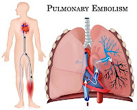
Your clinical Dx is pulmonary embolism.

Which one of the following would not be a feature of pulmonary embolism in this pt?
A. Dyspnoea
B. Tachypnoea
C. New-onset atrial fibrillation
D. Fever
E. Bradycardia

Answer: